
TL;DR

Prime made for students and young adults
- Fast, free delivery for dorm and study essentials
- Prime Video and Amazon Music included
- Member-only deals
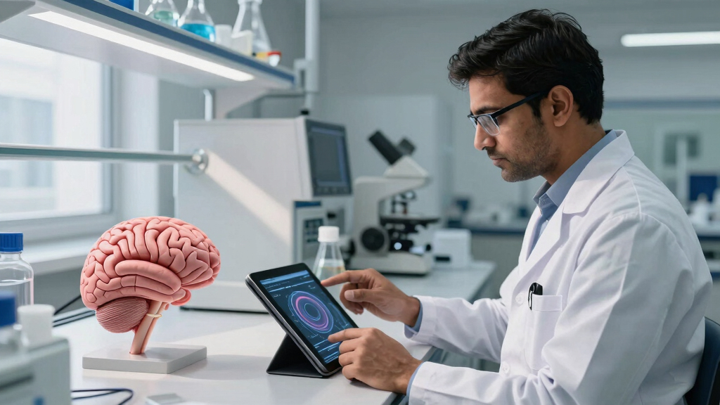
Indian scientists have produced the most detailed 3D atlas of the human brainstem to date. This development enhances understanding of brain anatomy and could impact neurological research and treatment.
Indian scientists have developed the most detailed 3D atlas of the human brainstem, a breakthrough in neuroanatomical mapping. This new atlas offers unprecedented resolution and detail, which could significantly impact neurological research and clinical practices worldwide.
The project was led by researchers at the Indian Institute of Neuroinformatics, who used advanced imaging techniques and computational modeling to create a comprehensive 3D map of the human brainstem. The atlas captures intricate structures, nuclei, and pathways with a level of detail not previously achieved in publicly available neuroanatomical resources.
According to the lead researcher, Dr. Anjali Mehta, the atlas was constructed using high-resolution MRI scans combined with histological data, allowing for precise delineation of brainstem regions. The team reports that this resource can aid in better understanding of brainstem functions and disorders.
Potential Impact on Neuroscience and Medicine
This development provides a detailed representation of the brainstem, which is involved in vital functions such as respiration, cardiovascular regulation, and consciousness. The atlas may support improved diagnosis and understanding of neurological conditions like stroke, Parkinson’s disease, and brainstem tumors. It can also serve as a reference for neurosurgical planning and research into brain connectivity and pathology.
As an affiliate, we earn on qualifying purchases.
Advances in Brain Mapping and Neuroanatomy
Previous brain atlases have primarily focused on the cerebral cortex, with less detailed representations of the brainstem due to its complex structure. While international efforts have produced some 3D models, none have matched the resolution achieved by this Indian team. The project builds on recent technological advances in imaging and computational modeling, which have facilitated detailed brain mapping.
The Indian team’s work aligns with global initiatives such as the Human Brain Project and the Allen Brain Atlas, but emphasizes the detailed mapping of the brainstem and its structures.
“This atlas provides a detailed view of the architecture of the human brainstem, which can support further research into neurological disorders.”
— Dr. Anjali Mehta
As an affiliate, we earn on qualifying purchases.
Remaining Questions About Atlas Validation and Accessibility
The extent of the atlas’s accessibility to the global scientific community and its validation against other models remain to be clarified. Peer review and independent validation are anticipated following publication.
Further investigation is needed to determine how the atlas might be integrated into clinical practice or personalized medicine approaches.
brainstem model for medical students
As an affiliate, we earn on qualifying purchases.
As an affiliate, we earn on qualifying purchases.
Next Steps: Validation, Publication, and Clinical Integration
The researchers plan to publish detailed findings in a peer-reviewed journal and make the associated data sets available to the scientific community. Validation studies comparing this atlas with existing models are expected to follow.
Efforts to incorporate the atlas into neurosurgical planning tools and neuroimaging software are also anticipated, which could support clinical applications.
As an affiliate, we earn on qualifying purchases.
Key Questions
How does this new atlas differ from previous brainstem maps?
The new atlas offers higher resolution and more detailed 3D visualization of brainstem structures compared to earlier models.
Will this atlas be available for global research and clinical use?
The team intends to publish the atlas and share the data with the scientific community, though specific access details are still being finalized.
What practical applications could this atlas have?
It may support improved understanding of brainstem functions, assist in diagnosing neurological disorders, and aid in planning neurosurgical procedures.
Are there any limitations or challenges remaining?
Further validation and integration into clinical tools are ongoing processes, and its utility in personalized medicine has yet to be established.
Source: hn
Halloween Picks
halloween
As an affiliate, we earn on qualifying purchases.